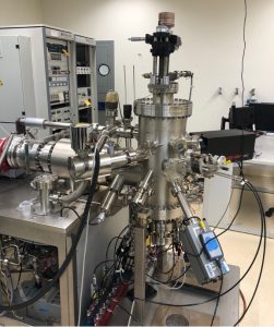
Thin Film Deposition
MBE system from Omicron/Scienta; six Kundsen cells with computer automated shutter control; oxygen plasma source (Mantis Deposition Inc.); reflection high energy electron diffraction (RHEED) system, equipped with a 20 kV electron gun (Staib Instruments) and digital camera to acquire videos and images; quartz crystal monitor and residual gas analyzer systems to measure beam flux and growth chamber chemical environment.
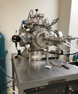
Kurt Lesker system with two sputter targets, quartz crystal monitor for rate measurement.
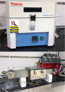
Post-Deposition Sample Processing
Two Thermo Scientific Lindberg/Blue M* Mini-Mite tube furnaces; one Thermocraft XST-2-0-12-mini-120 tube furnace for post-growth ozone and fluorination treatments; custom-built furnace for wafer-scale topochemical reactions.
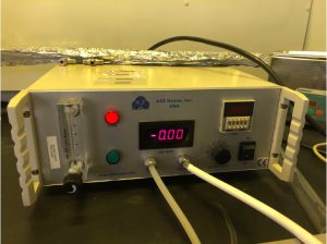
(A2Z Ozone Inc.) for ex situ, atmospheric pressure film oxidation
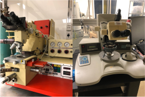
Mask aligner (Karl Suss MJB-3); custom-built spin-coater for photolithographic sample patterning; Wire bonder (Kulicke & Soffa KS4523A)
Materials Characterization
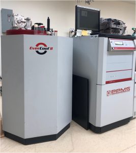
Quantum Design Physical Properties Measurement System (PPMS) with 1.8 to 400 K temperature range, magnetic field strength of up to 9 T, EverCool-II cryogen-free dewar. The system has the P400 option for the PPMS allowing for four-point resistivity and Hall effect measurements; high-resolution horizontal sample rotator with computer-controlled stepper motor (option D310B), allowing for magnetotransport measurements with full 360° in-plane and out-of-plane rotations; vibrating sample magnetometry (VSM) option allowing for dc magnetization measurements over the full temperature and field range of the system with a minimum resolution of 10-6 emu. External electronics include a Keithley model 6220 precision current source, model 2182A nanovoltmeter, model 2200 high voltage supply, model 2400 sourcemeter and model 248 power supply (< 5 V).
J.A. Woollam M2000U variable-angle spectroscopic ellipsometer with a heated stage capable of temperatures up to 300°C. Olympus BX51 optical microscope with digital camera and a Dell Optiplex workstation.
We also make extensive use of Drexel’s Materials Characterization Core facility, which has capabilities for x-ray diffraction, x-ray photoemission spectroscopy, and electron microscopy.